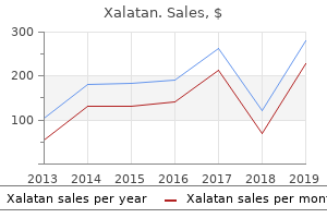
Cheap xalatan 2.5 ml with amex
Supine position allows access for both extension osteotomy and patellar advancement. Dorsal and plantar intermetatarsal ligaments provide further stability between the second through fifth metatarsal bases. Open reduction and internal fixation is generally mandatory to restore talar anatomy precisely. It is then reflected anteriorly off the intertrochanteric line, leaving the anterior hip capsule intact. Significant (external) derotation may sometimes be accompanied by visible bowing of the femur due to unmasking of the normal anterior bow. Shriners Hospitals for Crippled Children, Symposium on Caring for the Child with Myelomeningocele, American Academy of Orthopaedic Surgeons, 2002. The posterior tibial nerve and artery course posterior to the medial malleolus and split into the medial and lateral plantar nerves. Physical examination should include gross inspection of both lower extremities with the patient standing, walking, and sitting to determine the location of deformity as well as the alignment of adjacent structures (in particular the hindfoot and knee) that may contribute to perceived deformity as well as affect the surgical outcome. A formal synovectomy with sharp dissection is performed for removal of polyethylene wear particles and improved exposure. Avoiding fibular stabilization, however, does not convincingly decrease and perhaps even increases the chance of angular deformity. Vertebral anomalies cause scoliosis by an imbalance in bone growth, whether an increase on a side associated with a hemivertebrae, or retardation on the side associated with a vertebral bar. The proximal extent is the midpatella and the distal aspect is a few centimeters distal to the tibial tubercle fracture bed. A 4-mm half-pin is inserted bicortically into the base of the first and fifth metatarsals. The radial nerve enters the anterior compartment of the arm in the distal third of the upper arm and travels between the brachialis and brachioradialis over the anterolateral distal humerus before it enters the supinator muscle in the proximal forearm. The bilobed flap design must be drawn appropriately to take advantage of the redundant tissue on the ulnar side of the wrist. Next, using a blunt pin or straight instrument, mark on the skin the desired approach trajectory to the physeal bar. Cruciate ligament, which includes transfer ligaments, restrains against atlantoaxial anteroposterior translation. A 2001 study of 862 supracondylar fractures treated with open reduction found 55% excellent results, 24% good results, 9% fair results, and 12% poor results 5. Many cases can be managed nonoperatively, but orthopaedists need to maintain familiarity with operative techniques. A shallow trochlea groove, patella alta, patellar tilt, and a lateralized tibial tubercle can increase the risk of dislocation,5 along with a hypoplastic tibial tubercle and valgus knee alignment. The suture or wire tension band is anchored about a more proximal screw placed parallel to the articular surface. When arthroscopic evaluation and treatment of intraarticular disease is completed, the instruments are removed and the traction is released. Once the incision has been made, the entry point for the nail is identified 2 cm superior to the growth plate at the midpoint of the femur anteroposteriorly. Anterior Iliac Crest Graft In larger children, the anterior iliac crest can be used. Occasionally there may be large metaphyseal fragments that may be more appropriately stabilized with one or two lagged cancellous bone screws. If the joint aspiration is performed in the operating room, the arthrotomy can be performed in the same setting. To prevent subluxation and dislocation of an adjacent joint during limb lengthening: Correct hip instability before performing femur lengthening. This soft tissue tunnel is expanded down toward the medial epicondyle of the femur.
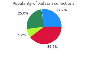
Discount xalatan 2.5 ml free shipping
Specifically, the surgeon must ensure the meniscus is not injured or interposed in the fracture before reduction. Stretching of the nerve with rapid distraction may result in nerve injury, and it may be necessary to decrease the rate of distraction or even stop distraction temporarily. In patients with cerebral palsy, the medical history correlates very strongly with postoperative complications. The lag screw can be placed from posterior to anterior with bicortical purchase achieved in each screw. Adson forceps identify the inguinal ligament with the external oblique fascia proximal and medial. For the older infant and child an anterior approach to the hip joint allows more extensile exposure. These patients often have a progressive deformity and are better treated at skeletal maturity using other fixation methods. The femoral osteotomy is just below the lesion, to provide disease-free bone stock for fixation of the proximal femur replacement. If a deficit is still present after reduction, a vascular study may be considered versus immediate operative exploration to evaluate for transient spasm or vascular injury. If patients return to activity before the cartilage has become firm, they will typically complain of pain with maneuvers such as squatting or jumping. Concern is increased if greater than 45 degrees of rotational malalignment is judged to be present. Approach A dorsolateral approach to the base of the fifth metatarsal is preferred. Hip, knee, ankle, and foot orthoses are a time-honored treatment and are used postoperatively in many centers. If bone quality is reduced, another screw can be inserted through the offset of the plate for additional stability. Evolution of the concept of an extensible nail accommodating to normal longitudinal bone growth: clinical considerations and implications. The labrum is squeezed between femoral neck and acetabular bone and eventually degenerates and ossifies. Adequate padding is applied over and beneath the pins to allow for support before cast immobilization. Looking specifically at elderly patients (older than 60 years), functional outcomes steadily improved over 1 year of follow-up, albeit at a slower rate than in the younger patients. The neurologic examination should include motor strength and a sensory examination of the upper and lower extremities. The child may have mild pain early or the pain may be so severe that the patient is unable to walk. Anatomical basis of variability in injuries of the medial malleolus and the deltoid ligament. The surgeon should push the rod to line up with the L5 lamina only and then twist the wires (we suggest a jet wire twister). This usually involves a small bend to accommodate both the proximal femur and the distal metaphysis. Preformed antibiotic-loaded polymethylmethacrylate spacers are commercially available, but the concentration of antibiotic in the cement is predetermined by the manufacturer. After complete release at the distal end of the A1 pulley, the cut ends should be pointing palmarly and not toward each other. They noted that patients with preoperative hindfoot valgus had improved alignment with varus overcorrection of the distal tibia and recommended overcorrection by 5 degrees in these patients. Cases of recurrence of triggering after surgical release have been attributed to incomplete release. A strip of Xeroform dressing can be wrapped around the pins, followed by fluff dressings.
Diseases
- Robinson Miller Bensimon syndrome
- Costocoracoid ligament congenitally short
- Acrorenal syndrome recessive
- Central nervous system protozoal infections
- Spondyloepiphyseal dysplasia
- Deafness peripheral neuropathy arterial disease
- Bhaskar Jagannathan syndrome
Proven xalatan 2.5 ml
Usually, the dummy axis pin can be slightly bent and the hinge axis reoriented to the knee rotational axis. Allogeneic cancellous bone graft and a Burch-Schneider ring for acetabular reconstruction in revision hip arthroplasty. Although seldom indicated, Leung and Tse described a lateral mini-plate buttress technique for the open physis. Persistent tightness of the hamstrings is associated with continued pain and, perhaps, an ultimate need for operative treatment. Gabriel and associates9 reported the results of 29 patients who underwent 56 physeal procedures with a percutaneous epiphysiodesis. Results of thoracoscopic instrumented fusion versus conventional posterior instrumented fusion in adolescent idiopathic scoliosis undergoing selective thoracic fusion. Complications have been reported while treating these complex fractures with other methods of fixation, such as titanium elastic nails or external fixation. The patient is allowed to bear weight to tolerance, and early progression to range of motion is encouraged. The gracilis and semitendinosus, found on the undersurface of the sartorius fascia, must be preserved. Care must be taken not to dissect excessively near the ulnar epiphysis, to prevent injury to the vascular supply to this area. Close attention is paid to the Canale image of the talar neck to be sure there is no malalignment of the neck fragment. Patients presenting with severe ankle pain and tenderness, hypoesthesia or anesthesia in the first dorsal web space, weakness of the extensor hallucis longus and extensor digitorum communis, and pain on passive stretch of the great toe may require release of the superior extensor retinaculum. Specific anatomy and growth plate closure patterns create certain fractures in adolescence. The bony landmarks and the location of the ulnar nerve are drawn carefully before the procedure to avoid inadvertent neurovascular injury. Milch type I fractures (the less common) traverse the metaphysis and physis as well as extend across the ossification center of the lateral condyle. The carcinomas that commonly metastasize to bone are those of the prostate, breast, kidney, thyroid, and lung. Cemented total or hemiarthroplasty for femoral head, neck, and peritrochanteric lesions remains, in general, the procedure of choice in this patient population, with good to excellent outcomes relative to the omnipresent comorbidities. Correction of this rotational deformity is crucial in restoring the medial column and the weight-bearing function of the first ray. The calcaneus has a shock absorber function by assisting in mobility of the ankle and subtalar joints, thus allowing the foot to accommodate to variations in terrain. Lengthening of the rectus femoris and hamstring muscles is recommended for positive muscle tightness. This analysis found no evidence of excessive early migration or loosening of the components. The drill is introduced through the sleeve, and the bone is drilled (both cortices). The reduction maneuver may be facilitated with a prior or concurrent closed reduction. The wire should be directed from distal lateral to proximal medial, penetrating the cortex medially. If knee range of movement must be limited for a period of time, a knee brace that allows movement only through a prescribed arc of motion may be necessary. The segmental arteries and veins originate from the aorta and vena cava, respectively, and traverse the vertebral body. Restoration of the joint line and the posterior condylar offset is crucial to the overall success of the revision process. The affected leg is draped separately in a bag to allow free movement of the hip joint.
Generic xalatan 2.5 ml with amex
The awl is temporarily left in its intraosseous position before insertion of the radial flexible intramedullary nail. Synovial white cell count differential may be of greater value, but data to recommend this routinely are limited. This can be aided by placement of a vise grip on the rod in the plane of the lordosis once the S-portion of the rod is positioned over the sacral ala. The intramedullary screw is placed as described for percutaneous intramedullary screw fixation. Impaction grafting and wire mesh for uncontained defects in revision knee arthroplasty. Progressively larger and more mature-appearing lesions with ossification are seen on the surface of the bone as the distance from the physis increases, so they appear to be migrating into the diaphysis of long bones. The procedure lasted a mean of 36 minutes, and in all patients a physeal closure developed. If difficulty is encountered, soft tissue can be peeled off the lateral border of the patella to make it more mobile, and any osteophytes that are present can be removed. The originators of this technique suggest nail removal by about the sixth postoperative month. Nonetheless, the success rate increased to 94% after a repeat surgery, reaching a 100% healing rate in patients who underwent more than two repeat surgeries. The Foley catheter is removed on postoperative day 2 and urinary function is closely monitored. The authors stated that metal wedge augmentation for tibial bone deficiency is a useful option. In younger children, polytrauma, head injury, high-energy trauma, an open fracture, severe comminution, or body habitus incompatible with spica cast care are relative indications for operative management. Nail plate deformity is common after simple complete syndactyly release in the presence of a synonychia. Type 2B defects are similar to type 2A defects, with the exception of loss of the superior rim. The posterior edge of the gluteus medius is retracted anteriorly to reveal the piriformis tendon. This extra-articular ligament is tensioned with the knee in 90 degrees of flexion to prevent an extension contracture. Again, the clamp bolt should be loose enough to allow translation of the soft tissue cover through the arm. An osteotomy is made parallel to the ankle joint, and an opening wedge correction is performed and filled with bone graft. Patients with low-grade dysplastic spondylolisthesis are at greater risk for progression, development of neurologic deficit, and need for operative intervention. If there is friction during the drop-leg test, the hinge and knee rotation axis needs to be examined and adjusted. Laterally, very dense cortical bone along the proximal neck presents an excellent extra-articular location for advancing a second interfragmentary screw. Before draping the patient, we place a marker over the hemivertebra region and obtain a radiograph. Proximal hamstring lengthenings during adolescence also run an increased risk of developing heterotopic ossification in the proximal muscle release site. Evidence of talar dome collapse presented in half of these cases by 14 months after the injury. Inadequate exposure with continued forceful retraction of the extensor mechanism risks avulsion of the patellar ligament from the tibial tubercle. The fixation pins should not be inserted near the anterosuperior iliac spine point, because this point is required for pelvic registration.
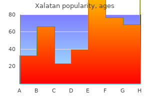
Buy cheap xalatan 2.5 ml line
Important risk factors for operative treatment complications include smoking, diabetes, peripheral vascular disease, and steroid use. Range-of-motion exercises Follow-up radiographs through resorption and reconstitution phases prior to resumption of sport-specific exercises. Immediate postoperative treatment requires application of sterile ankle dressings and a well-padded, short-leg dressing with a posterior plaster splint. The Risser sign should be evaluated by assessing the ossification of the iliac apophysis, giving it a grade between 0 and 5. Small lesions (ie, those that involve less than one third to half of the bone width) in weight-bearing bones that have low risk for fracture can also be observed. The infection has likely been present since the original procedure, but because of the low virulence of the infecting organism, classic signs of infection are lacking, and hip pain may be the only presenting symptoms. In older children with significant deformity, a bipolar release is the first step. A bone hook can be used to apply a direct lateral force to disimpact the femoral neck fracture. A bone hook is placed around the neck of the femur anteriorly, and the leg is externally rotated to allow for dislocation of the hip, ie, the hip is placed in the figure-4 position. This cut should be made at right angles to the anatomic axis of the tibia in the coronal plane. Positioning the positioning for knee fusion is the same for each surgical approach. Replace wires with cancellous screws following insertion of the definitive implant. After the proximal half-pins have been placed, the rail is disengaged from the proximal clamp. The synovial fluid should be evaluated and cultured for the presence of microoorganisms. If the bone is strong enough, no pain should occur with dynamization in the office setting. Lower extremity positioning with internal and external rotation greatly enhances the extent of visualization and allows safe recontouring under direct vision. The acetabulum typically demonstrates 15 to 20 degrees of anteversion, as does the femoral neck. Nineteen percent of the patients experienced a clicking, locking, or clunking noise or feeling in the first 6 months after surgery, but it was painless and disappeared gradually. The data that show 20% coexistence of developmental dysplasia of the hip support the theory of intrauterine malposition and crowding. Although all are part of the same muscle group, their structure and function differ. After insertion of the tibial rod, the wire is then drilled into the proximal fibula. There is no examination under anesthesia to determine the amount of varusization to accomplish. If it is a nondisplaced fracture or the separation is minimal, a contralateral comparison radiograph may help confirm the diagnosis. After release of radial tethering tissue and rotation of flaps, the skin is sutured. Arthrodesis of the fourth and fifth tarsometatarsal joints should be avoided if possible. Position of pin for percutaneous pin-assisted reduction of a displaced radial neck fracture. Broaching is complete when the broach ceases to advance, the pitch of impaction increases, and good cortical contact is obtained. Despite initial fluoroscopic views indicating an adequate reduction, the radiographs indicate a poor reduction.
Xalatan 2.5ml overnight delivery
Patellar component position Thickness of resurfaced patella Medial or central positioning of the patellar component is acceptable. For patients with external fixators, pin tract infections are likely to occur and are initially treated with orally administered antibiotics. This more accurate and precise positioning and alignment of the components should reduce the rate of long-term complications and revisions. The ossification center of the capitellum appears at 18 months and completely fuses by age 14. If necessary, the fibular osteotomy is performed using a separate 2-cm lateral incision that parallels the fibula and is centered over the point of the osteotomy. Rett Syndrome this is an X-linked disorder that affects females almost exclusively. Sonography of the spinal cord is helpful in infants younger than about 4 months of age who have congenital spine anomalies. At 8 weeks noncontact sports were allowed, and return to full activity was possible at 12 weeks after surgery. Neuromuscular patients with a migration index less than 25% can be observed as long as their abduction remains greater than 45 degrees. The surgeon and anesthesiologist should plan for the possibility of large intraoperative blood loss. Extension of the incision on the opposite side of the displacement of the distal fragment allows for removal of soft tissue obstacles to reduction. Most commonly preferred procedures include unipolar release, bipolar release with or without Z-plasty lengthening of the sternal head, and the extended procedure for older children and resistant cases. Preoperative Planning Preoperatively, a careful neurovascular examination should be performed and documented. Fractures of the posterior body of the talus are performed through posteromedial or posterolateral surgical approaches. Elbow stability should be checked and full range of motion confirmed before closure. We occasionally suture the abductors to the vastus lateralis, the tensor fascia lata, or the host greater trochanter, if available. Stems are used routinely, both to support the revision component and to assist with alignment. Alternatively, the surgeon can hold his or her thumb on the proximal fragment and push downward while an assistant applies traction to the forearm with the elbow flexed at 90 degrees. Fracture is treated with reapplication of the frame, casting, intramedullary rod fixation, or plate application. Infantile tibia vara is most prevalent in African-American females and is associated with obesity, internal tibial torsion, and leg-length discrepancy. The broad insertion of the medial collateral ligament presents an issue only for severe bone defect unless it is found to be incompetent on the preoperative physical examination. The chest tube may be removed when drainage is less than 80 mL over 12 hours and serous color returns (with good pleural closure, removal usually is done on the first day). The acetabulum is palpated (if done with an open reduction) or the hip taken through full range of motion (if done as an isolated procedure) to ensure pins are extraarticular. True scanograms use a slit beam that is perpendicular to the patient that scans the length of the limb and therefore has no magnification. In older children and teenagers with a longstanding jump or crouch gait pattern, lengthening of the hamstring muscles alone may not result in complete knee extension when the hip is placed in extension. The incision is located over the anterior compartment, lateral of the palpable crest of the tibia and curving gently medially at the ankle joint. Necrotic bone usually is yellowish, because it does not bleed, so the margin between necrotic bone and living bone is well recognized. The stance and swing phases of each cycle are separated by the dashed black lines. A small or comminuted superomedial fragment makes rigid fixation harder to achieve and may call for alternative techniques. It is very important when placing the proximal femoral pins to ensure that they are not positioned too anteriorly in the femoral shaft. This is a limited approach, not the wide extensile exposure needed for open reduction and internal fixation.
Quebrachol (Beta-Sitosterol). Xalatan.
- Burns, prostate infections, sexual dysfunction, preventing colon cancer, rheumatoid arthritis, psoriasis, allergies, cervical cancer, fibromyalgia, systemic lupus erythematosus (SLE), asthma, baldness, migraines, chronic fatigue syndrome, menopause, and other conditions.
- What is Beta-sitosterol?
- Trouble urinating from an enlarged prostate, or "benign prostatic hyperplasia" (BPH).
- Are there any interactions with medications?
- Are there safety concerns?
- High cholesterol.
Source: http://www.rxlist.com/script/main/art.asp?articlekey=96902
Buy 2.5ml xalatan mastercard
Tornetta prefers the lateral position, with conversion to the extensile lateral approach if percutaneous manipulations are unsuccessful. The goal of revision component augmentation is stable contact between metal and host bone without resorting to a custom implant. Patients receiving total femur replacement may require the use of continuous passive motion machines for rehabilitation of the knee replacement. Note the 90-degree-flexed position of the elbow and the 90-degree external rotation of the shoulder. The foot is further plantarflexed to expose the posterior ankle joint, which is released, exposing the posterior calcaneus and the Achilles tendon. A 13-year-old with myelokyphosis with diastasis beginning at T6 with 127 degrees of kyphosis. It also eliminates the potential for fractures around the pins, because they are not placed in diaphyseal bone. The interphalangeal joint typically has full passive range of motion, while the metacarpophalangeal joint is fixed in flexion, differentiating congenital clasped thumb from pediatric trigger thumb. The turndown group had a higher increase in arc of motion than the osteotomy group, but they also had a higher degree of extension lag. A synovial white cell count of greater than 2000 cells/mL or over 65% polymorphonuclear leukocytes is suggestive of infection. The main advantage of this technique is that it allows for restoration of bone stock. The posterior approach is a standard posterior midline incision with subperiosteal dissection out to the tips of the transverse processes. Options available for dealing with severe femoral bone loss include the use of a long cemented stem or press-fit stems, impaction allografting, resection arthroplasty, allograft prosthetic replacement, and modular replacement. Research attempting to demonstrate that motion occurs within a bipolar prosthesis has yielded conflicting results. Initially the wire is gently pushed through the soft tissue until it hits bone cortex. Grissel syndrome: torticollis associated with retropharyngeal abscess or post-tonsillectomy status. Densely adherent skin is much harder to close and may represent a higher risk for necrosis. Consequently, in the revision setting where posterior stabilized or "super stabilized" or "total stabilized" type components are used, additional anterior resection of the tibial plateau may be required to restore neutral slope. The U-shaped seating chisel is inserted into the cortical windows with the direction defined by the two K-wires. Since such films are poorly tolerated in an awake child, they are rarely obtained outside of the operating room. This is usually necessary to avoid distraction by an intact fibula after resection of the pseudarthrosis. Patients who are symptomatic at initial presentation are often at risk for progressive neurologic symptoms. In patients who have had previous hip surgery, the sciatic nerve may be entrapped within scar tissue. Surgical correction is indicated when the child is not tolerating seating with a combination of either seating adjustments or a soft orthosis. Pain usually is recognized along the medial joint line, but its localization is unreliable. In children and adolescents, this most commonly occurs in the presence of a spondylolytic defect or a nonunion of the pars interarticularis. The osteotomy rotates the acetabulum to improve anterior and lateral femoral head coverage. The surgeon should avoid placing the pins in the region of the axillary and musculocutaneous nerve. Sternocleidomastoid pseudotumor and congenital muscular torticollis in infants: a prospective study of 510 cases. Patient outcome with reinfection following reimplantation for the infected total knee arthroplasty.
Buy generic xalatan 2.5ml online
The surgeon should incise slightly lateral to the fat on the fascia of the tensor. Flynn and associates6 reviewed their first 50 cases and found that insertion site irritation was the most common problem encountered (18% of cases). The patient is assessed every 2 weeks in the outpatient clinic with radiographic and clinical examinations. Use of medial and lateral augments simultaneously should be an indication to carefully evaluate possible elevation of the joint line. A wedge of bone is resected, apex medial in valgus ankles and apex lateral in varus ankles. The thickness of the bearing should be such as to restore the ligaments to their natural tension so that when a valgus force is applied to the knee, the artificial joint surfaces distract a millimeter or two. An oscillating saw is less likely to cause uncontrolled fracture in the ilium cut. The knee should be able to flex 60 degrees at this point without excessive tension or disruption of the repair. The clinician should determine whether the patient has any other complaints of pain beyond the forearm shaft region (eg, wrist or elbow tenderness). Preoperative measurements from plain films provide a reliable guide for the length and width needed. Currently, the author uses somatosensory evoked potentials and transcranial electrical motor evoked potentials to evaluate the brachial plexus nerve function during surgery. The prosthesis should remain at or lateral to the medial floor of the acetabulum, and the superolateral corner of the component should fall near the superolateral border of the acetabulum. The shape of the tibial articular surface is concave, with distal extension of the anterior and posterior lips. Early passive range of motion is not recommended because of the risk of heterotopic ossification. Now the free ends of the looped wire are brought down over the graft and passed below the spinous process. No matter what procedure is used for treating the radial dysplasia, the patients all have a high incidence of recurrent deformity as they get older. Patients with pain without instability should not be operated on unless all else fails. The hip is internally rotated until the femoral head trial is coplanar with the rim of the acetabular component. During the distraction phase, physical therapy is continued daily, with formal therapy occurring 5 days per week. This is accomplished by sequential assembly of modules that are then connected by a spanning carbon rod. Tanner staging should be confirmed at the time of surgery after the induction of general anesthesia. Many children have tendon transfers as young as 7 years old; with continued skeletal growth, they may have recurrent deformity. A proximal osteotomy can be used for deformity correction with a concurrent distal osteotomy for lengthening. This is because their hips can rotate internally more than they can rotate externally. Straight leg raise should be done to test for nerve root compression or hamstring tightness. Early mobilization, weight bearing, and strengthening are encouraged to avoid knee stiffness. Iatrogenic fractures of the distal femur, proximal tibia, or both can occur with casting and manipulation. The gastrocnemius can be selectively released or lengthened either at its origin, which is uncommonly done, or the tendon of the gastrocnemius, proximal to the conjoined tendon.
Purchase 2.5ml xalatan
Routine radiographic screening studies in search of early metastatic disease are not very helpful. If one is to err, one would prefer too little tension than too much, as the transfer tends to tighten over time, particularly if performed in a young child with significant remaining growth potential. If any contouring is necessary in the region where the cranial and caudal rods meet, closed dual connectors must be used, with an overlap of 2 to 4 inches to allow future lengthening. Seventy percent of patients who are ambulatory retain their ability to ambulate after radiation therapy to the lower extremities. Contraindications for surgical reconstruction include: Infection Inability to comply with postoperative immobilization and the physical therapy program For these rare instances, cast or brace immobilization in full extension for 6 to 8 weeks followed by a physical therapy program to regain motion and strength may be appropriate. The gray area represents the optional trochanteric osteotomy, which is recommended only when the correction angle exceeds 25 degrees. Multiple extra-articular drill holes (three or four) are placed in the patella to create transosseous suture tunnels for imbrication. About 45% of all primary tumors will be detected in the lung on the chest radiograph. A spica cast is frequently used if the radiograph shows laxity and lateral displacement, especially in the infant. The surgeon must maintain vigilance throughout the postoperative period for late development. Normal anteversion at birth is 45 degrees, decreasing to 10 degrees in boys and 15 degrees in girls by 8 years of age. If the fibula is an impediment to correction (it usually is not) an oblique osteotomy of the fibula is made and if necessary fixed with a plate and screws. Occasionally, test results may be equivocal, in which case nuclear medicine imaging is required to confirm absence of infection. In the isthmic type of spondylolisthesis, secondary to a pars defect, the high shear and compressive forces occurring through the lumbar spine and lumbosacral joint are less well resisted. With reduction maintained, the screw is gradually advanced under fluoroscopic guidance, making sure that the growth plate is not traversed. Proximally to the osteotomized site, the periosteum on the medial edge of the iliac crest is incised and reflected medially with the origin of the iliacus muscle. Uncemented fixation into the distal femur may be an option with some reconstruction systems. Isolated palsy has been reported secondary to constrictive dressings and after proximal ulna fracture. The medial and lateral epicondyles have been screwed into their corresponding anatomic locations on the bulk allograft. The knee examination is repeated under anesthesia, including ligamentous testing, range of motion, and the McMurray test to evaluate whether significant lateral meniscal instability is present. The periosteum is incised with a sharp knife and is elevated circumferentially with a periosteal elevator. Multiple Kirschner wires and carefully placed pointed clamps may helpful in gaining and maintaining reduction. In our experience, compliance with activity modification and brace use and effectiveness limits the success of this treatment. The ligamentum teres originates from the transverse ligament over the acetabular notch and inserts into the fovea of the femoral head. Use of a total-contact orthosis is essential throughout growth to reduce stress through the pseudarthrosis and maintain consolidation. The patient and surgeon should discuss the use of auto- or allograft material for bone grafting of the osteotomy site.
Buy xalatan visa
Once stabilized, the articular segment can then be attached to the tibial diaphysis through open or minimally invasive plating (or external fixation). Controversy exists regarding the exact numbers, however, with reported acceptable angulation ranging from 20 to 60 degrees. Female nail appears to be protruding but is actually deep to the articular cartilage and is not causing impingement. These screws are attached to the inner combination clamps and the bolts are tightened. Preoperative evaluation of the genitourinary system with a screening ultrasound and evaluation of the cardiac system with an echocardiogram are necessary if these have not been performed, given the rate of anomalies associated with congenital scoliosis. The risk of redislocation is three times higher in patients treated with immediate mobilization versus immobilization with cast or brace. Straight ream proximally to the same diameter as the minor diameter of the implant. The subcuticular closure is performed in layers, and a cast is placed with the foot in neutral or slight dorsiflexion. In classic hemihypertrophy, upper extremity hypertrophy as well as hemifacial asymmetry may be present. Surgery for cerebral palsy, part 3-classification and operative procedures for thumb deformities. Delayed complications most commonly include osteolysis, aseptic loosening, and late septic prosthetic arthropathy. External oblique fascia is divided along the inguinal ligament and retracted by Army-Navy retractors to visualize the internal oblique. Kirschner wires provide provisional fixation, and a plate is contoured to the superior calcaneus, behind the posterior facet. If residual acetabular dysplasia exists, a Salter innominate osteotomy can be performed at that time. When performing radial head excision, the surgeon has to be careful in the initial dissection to avoid injury to the posterior interosseous nerve as well as to the stabilizing structures of the elbow. Clear adhesive surgical drapes (3M Steri-Drape Towel Drapes) are placed around the perimeter of the surgical site, extending from the hairline to the top of the gluteal crease (regardless of levels to be fused, the entire spine should be draped). There are growth plates beneath the capital femoral epiphysis, the greater trochanteric apophysis, and the lesser trochanteric apophysis. Gradually revascularization of the epiphysis begins, usually at the anterolateral area of the epiphysis. Complete exposure of the peripheral rim with subperiosteal dissection is performed to avoid injury to surrounding neurovascular structures such as the superior gluteal nerve and vessels. Fluoroscopic imaging is often necessary when placing screws at the L5 level because of the distorted anatomy. The surgeon is applying pressure to reduce the apex anterior angulation, while maintaining traction with the left hand. The ideal fulcrum is near the medial cortex of the tibia for maximal angular correction. This approach has the advantage of preserving the posterior retinacular vessels, which reduces the possibility that an iatrogenic avascular state will develop postoperatively in the remaining femoral head. The host bone is reamed to expose healthy, bleeding cancellous bone, including removal of all fibrous tissue and cement. It is important to appreciate the continuity of the tubercle and proximal tibial growth plates. Nonoperative treatment consists of activity modifications, anti-inflammatory medications, and swelling control (ice, elevation, and compression). The goal of reduction is to achieve a congruent articular surface without any step-off. The rectus femoris tendon is delivered into the medial incision (solid circle), where it will be transferred to the distal portion of the semitendinosus muscle tendon (dashed circle).
